Chongfan Technology
News
22
2026
-
06
The Xi’an Institute of Optics and Precision Mechanics of the Chinese Academy of Sciences has achieved new advances in the field of scattering microscopy.
Author:
Recently, the Transient Optics Laboratory of the Xi’an Institute of Optics and Precision Mechanics, Chinese Academy of Sciences, has made new advances in optical microscopy through scattering media. The research findings have been published in the Journal of Physics: Photonics. The paper’s co‑first authors are Associate Researcher Peng Tong and Assistant Researcher Li Runze, both from the Xi’an Institute of Optics and Precision Mechanics, while the corresponding authors are Researchers Dan Dan and Yao Baoli, also from the same institute.
Optical microscopy, with its unique advantages of non‑invasiveness and non‑destructiveness, is a cornerstone tool for probing microscopic biological processes. However, the refractive index within biological tissues is highly heterogeneous, leading to severe light scattering and wavefront distortion as photons traverse these structures—much like observing the microscopic world through multiple layers of frosted glass. This not only blurs images but also severely limits imaging depth. Current scattering‑compensation techniques typically suffer from system complexity, high computational demands, and reliance on labeling. Conventional transmission microscopy struggles to simultaneously correct wavefront distortions in both the illumination and detection paths, while wavefront shaping, though capable of focusing light fields through scattering media, is constrained by relatively large focal spot sizes, thereby limiting spatial resolution.
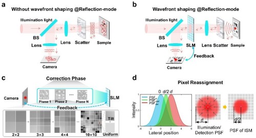
Figure 1. Principle of high-resolution imaging through multilayer scattering media via wavefront shaping and image-scanning microscopy.
In response, the research team innovatively developed a guide-star‑free, bidirectional wavefront‑corrected imaging technique. This approach uses the sample’s reflected light as the feedback signal and, with a single spatial light modulator, simultaneously compensates for wavefront distortions in both the incident illumination path and the returning detection path, thereby achieving noninvasive optical refocusing without the need for fluorescent labeling.
This technology integrates wavefront shaping with image-scanning microscopy, further reducing the size of the focused spot on top of scattering‑induced focusing. By employing a correction strategy that progressively increases the number of control elements and a hierarchical scattering compensation mechanism, it significantly enhances computational efficiency while maintaining high precision.
Experimental results demonstrate that this technique achieves high‑quality focusing in a three‑layer scattering medium with a total optical thickness of 1.82. Furthermore, by integrating pixel redistribution with deconvolution, the system’s resolution is improved by approximately 1.76‑fold compared to scattering correction alone.
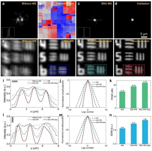
Figure 2. High-resolution imaging results through a scattering medium.
This technology addresses the longstanding challenge of simultaneously compensating for scattering-induced distortions in both the illumination and detection paths of optical microscopy, opening up new avenues for high-resolution imaging of deep‑lying biological tissues and holding promise for applications in biomedical diagnostics and other fields.
Peng Tong explained, “Light scattering by biological tissues is the primary obstacle that limits optical microscopy from achieving deep‑tissue, high‑resolution imaging. By precisely controlling the light field, we restore order to scattered light, effectively overcoming interference along the light‑propagation path and achieving a breakthrough—from ‘invisible’ to ‘visible’ and ultimately to ‘clearly visible.’”
This research was supported by projects including the National Natural Science Foundation of China, the National Key R&D Program, and the Shaanxi Provincial Key R&D Program.
In recent years, the research team led by Researcher Yao Baoli at the Xi’an Institute of Optics and Precision Mechanics has conducted in-depth studies and achieved a series of breakthroughs in the fields of imaging through scattering media and optical microscopy, including imaging through dynamic scattering media, non-invasive microscopy through scattering media, and two-photon adaptive optics imaging. Relevant findings have been published in journals such as Nanophotonics, Photonics Research, Applied Physics Letters, and Optics Express.
Source: Xi’an Institute of Optics and Precision Mechanics
LATEST NEWS
Thank you for visiting the official website of Chongfan Technology. If you have cooperation intentions or suggestions, please contact us through the following methods, and we will reply as soon as possible, thank you!
Address: Room 403, Building 6, Phase III of R&D, No. 36 Xiyong Avenue, High tech Zone, Chongqing, China.
Telephone: +86-13658337211
E-mail: Sales@cfkeji.net
Website: www.cfkeji.net
Mobile Version