
Chongfan Technology
News
28
2026
-
05
The Xi’an Institute of Optics and Precision Mechanics of the Chinese Academy of Sciences has achieved a series of significant advances in the field of optical microscopy.
Author:
In the microscopic realm, the transient response of cells to laser irradiation, the faint signals in deep tissues that are obscured by scattering, and the phase information in transparent samples—often difficult to observe directly—are all concealed within the subtle processes of light–matter interactions.
How to “chase light, capture speed, and discern the microscopic”—enabling microscopic imaging to keep pace with rapid dynamic processes, penetrate complex tissues, and retrieve hidden phase information—is a major challenge in advanced optical imaging and one of the key research priorities at the Xi’an Institute of Optics and Precision Mechanics, Chinese Academy of Sciences.
Recently, the research team led by Researcher Yao Baoli at the Xi’an Institute of Optics and Precision Mechanics, Chinese Academy of Sciences, has been tackling these critical challenges. They have achieved a series of breakthroughs in areas such as high-fidelity compressed ultrafast imaging, multi‑guide star–based parallel wavefront sensing, and dual‑encoding computational microscopy—making transient processes clearer, deep‑tissue imaging more precise, and lensless phase reconstruction more reliable. These advances have opened new avenues for observing biological systems, micro‑ and nano‑fabrication, and ultrafast physical phenomena.
Relevant research findings have been consecutively published in Optics Express, Optics and Lasers in Engineering, and Optics Letters.
Advancement 1: High-fidelity compressed ultrafast imaging for capturing cellular transient dynamics
To address the challenges of limited light throughput and reconstruction distortion in high-speed optical microscopy, the research team has innovatively developed a High-Fidelity Compressed High-Speed Imaging (HF-CHI) method. By integrating a high-transmittance static encoding mask, a dual-path synchronous acquisition architecture, and a multi-prior physical‑enhanced neural network (mPEN) developed by the team, this approach achieves superior spatial resolution—exceeding 181 line pairs per millimeter—at an equivalent frame rate of 50 kfps.
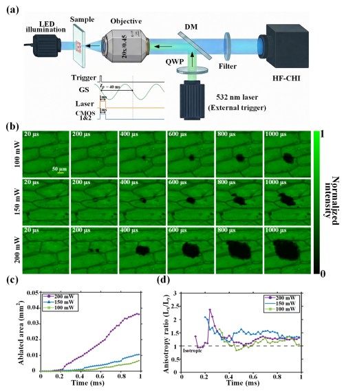
Figure 1. Photothermal ablation dynamics imaging results of onion epidermal cells.
This method has successfully resolved complex, non-repetitive transient dynamical processes such as cellular photothermal ablation, providing a powerful tool for research in biomedicine, micro‑ and nanofabrication, and ultrafast physics.
The relevant research findings, titled “High-fidelity compressed high-speed imaging for resolving rapid micro-dynamics,” were published in Optics Express in April 2026. The co-first authors of the paper are Dr. Li Xing and Dr. Wang Siying from the Xi’an Institute of Optics and Precision Mechanics, while the corresponding authors are Researcher Bai Chen and Researcher Yao Baoli, both from the same institute.
Paper link: https://doi.org/10.1364/OE.595532
Progress Two: Multi‑channel star‑based parallel wavefront sensing to enhance deep‑tissue imaging quality.
In deep-tissue biological imaging, spatially varying wavefront distortions induced by the sample severely degrade image quality. Conventional pupil‑plane adaptive optics (PAO) approaches struggle to achieve effective correction over large fields of view, while existing techniques based on multi‑guide star wavefront sensing are limited by the number of guide stars—typically only a few dozen.
In response, the research team has proposed a multi‑guide‑star parallel wavefront sensing (PWS) method that, without significantly increasing computational overhead or requiring enhanced hardware capabilities, can simultaneously leverage hundreds of guide stars to efficiently correct highly spatially varying wavefront distortions.
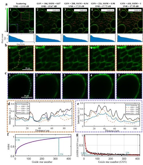
Figure 2: Comparison of corrected images under different numbers of guide stars.
This method significantly increases the number of available guide stars, offering a novel and highly efficient solution for mitigating complex wavefront distortions in deep-tissue imaging.
The relevant research findings, titled “Parallel wavefront sensing with hundreds of guide stars for anisoplanatic correction in two-photon microscopy,” were published in the May 2026 issue of Optics and Lasers in Engineering. The first author is Dr. Yang Ruiwen from the Xi’an Institute of Optics and Precision Mechanics, while the corresponding authors are Senior Experimentalist Yang Yanlong and Researcher Yao Baoli, both from the same institute.
Paper link: https://doi.org/10.1016/j.optlaseng.2026.109818
Progress Three: Dual-encoding computational microscopy overcomes the challenge of low-frequency phase loss.
Conventional lensless coded ptychography (CP) and similar methods commonly suffer from the loss of low-frequency phase information.
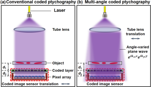
Figure 3: Comparison of the system principles between conventional coded stacked imaging and the proposed method.
The research team has proposed a computational microscopy imaging method based on multi-angle illumination and coded piling-up, termed Multi-angle CP. This approach employs a dual‑encoding scheme of “illumination–detection”:
·The illumination end generates plane waves with varying angles of incidence via lateral translation of the cylindrical lens, thereby providing phase diversity;
·A coding layer is introduced at the detection end to serve as a support-domain constraint, thereby enhancing the convergence of phase recovery. This strategy effectively improves phase reconstruction accuracy and overcomes the bottleneck posed by the loss of low-frequency phase information.
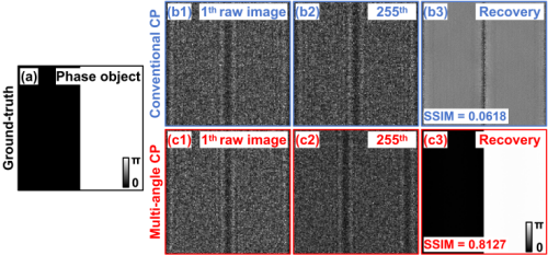
Figure 4: Comparison of simulation results between conventional coded stacked imaging and the proposed method for low-frequency phase targets.
This method effectively enhances the accuracy of phase reconstruction and addresses the challenge of missing low-frequency phase information.
The relevant research findings were published in the April 2026 issue of Optics Letters under the title “Lensless quantitative microscopy based on multi-angle illumination and coded ptychographic phase retrieval.” The first author is Yang Liming, a Special Research Assistant at the Xi’an Institute of Optics and Precision Mechanics, while the corresponding authors are Researchers Wu Tengfei and Yao Baoli, also from the same institute.
Paper link: https://doi.org/10.1364/OL.585444
The aforementioned research was supported by projects including the National Natural Science Foundation of China and the National Key R&D Program.
In recent years, Researcher Yao Baoli’s team has focused on tackling key challenges in advanced optical microscopy, developing a variety of novel optical imaging techniques. By further integrating artificial intelligence algorithms, the team has significantly enhanced the imaging capabilities, information‑acquisition dimensions, and performance metrics of these advanced optical microscopy methods. Relevant research has been published in journals such as Opto‑Electronic Advances, PhotoniX, and Ultrafast Science. The team has been granted multiple national invention patents and has received numerous awards and honors, including the First‑ and Second‑Class Awards for Science and Technology of Shaanxi Province, as well as recognition as a Key Innovation Team in Science and Technology of Shaanxi Province.
Source: Xi’an Institute of Optics and Precision Mechanics
LATEST NEWS
Thank you for visiting the official website of Chongfan Technology. If you have cooperation intentions or suggestions, please contact us through the following methods, and we will reply as soon as possible, thank you!
Address: Room 403, Building 6, Phase III of R&D, No. 36 Xiyong Avenue, High tech Zone, Chongqing, China.
Telephone: +86-13658337211
E-mail: Sales@cfkeji.net
Website: www.cfkeji.net
Mobile Version