
Chongfan Technology
News
28
2026
-
05
Biological Imaging—Cryo-EM + Mass Spectrometry, FIB-SIMS | Nature Methods
Author:
Cryo‑electron microscopy (cryo‑EM) enables high‑resolution structural characterization of biological samples, but it faces challenges in identifying their chemical composition.
Recently, Hannah Ochner, Tanmay A. M. Bharat, and colleagues at the MRC Laboratory of Molecular Biology at the University of Cambridge published in Nature Methods a method that integrates cryo‑electron microscopy with chemical imaging based on focused ion beam secondary ion mass spectrometry, enabling integrated spatiotemporal chemical analysis of unlabelled samples. This integrated workflow achieves subcellular localization of molecules within bacterial cells and is compatible with cryo‑light microscopy as well as focused ion beam thinning of eukaryotic samples.
A workflow‑driven biological discovery investigated the uptake of the ubiquitous chemical pollutant bisphenol AF by environmental bacteria. The study demonstrated that this compound accumulates within phase-separated condensates in the cytoplasm of exposed cells, and that even when bacterial efflux mechanisms are markedly upregulated, these pollutants cannot be effectively removed. Consequently, cryo‑electron microscopy combined with focused ion beam secondary ion mass spectrometry holds promise as an effective approach for mapping elemental and molecular features in near‑native biological samples.
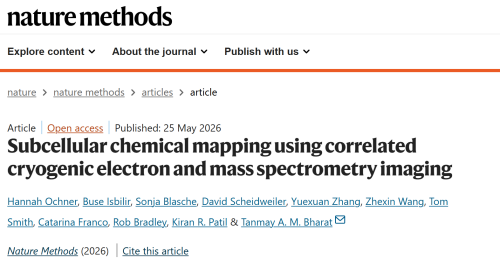
Subcellular chemical mapping using correlated cryogenic electron and mass spectrometry imaging. Subcellular chemical mapping based on correlative cryo-electron microscopy and mass spectrometry imaging.
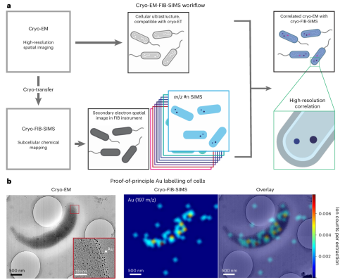
Figure 1: Workflow of cryo‑electron microscopy–focused ion beam secondary ion mass spectrometry.
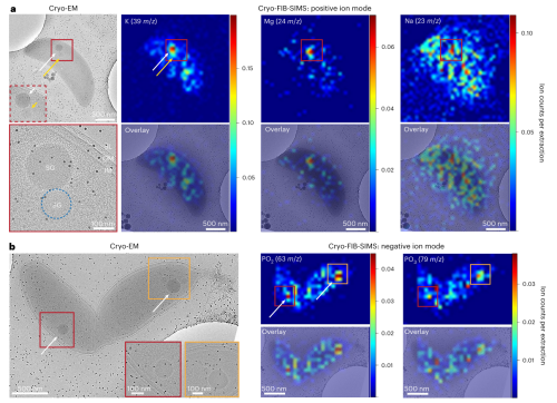
Figure 2: Correlative cryo‑electron microscopy–secondary ion mass spectrometry imaging at subcellular resolution in bacteria.
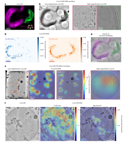
Figure 3: Workflow of cryo‑electron microscopy, focused ion beam secondary ion mass spectrometry, and optical microscopy, along with their compatibility for imaging thick samples.
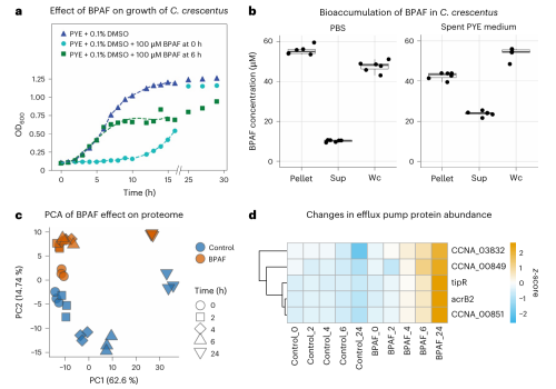
Figure 4: Effects of bisphenol AF on Caulobacter cells.
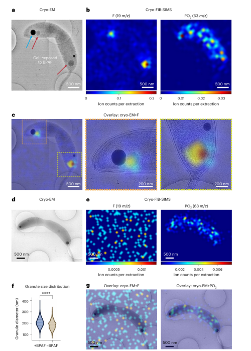
Figure 5: Localization of bisphenol AF within the cells of Rhodobacter sphaeroides.
Source: Today’s New Materials
LATEST NEWS
Thank you for visiting the official website of Chongfan Technology. If you have cooperation intentions or suggestions, please contact us through the following methods, and we will reply as soon as possible, thank you!
Address: Room 403, Building 6, Phase III of R&D, No. 36 Xiyong Avenue, High tech Zone, Chongqing, China.
Telephone: +86-13658337211
E-mail: Sales@cfkeji.net
Website: www.cfkeji.net
Mobile Version