
16

2024

-

09

Tsinghua University Xiao Bailong/Peking University Ouyang Kunfu Collaboration Reveals Key Phosphorylation Sites That Regulate Piezo1 Mechanical Sensitivity and Mechanotransduction Function in Vivo

Author:


Piezo1 is a mechanically activated cation channel that can convert mechanical forces into various physiological processes.Due to its large protein size of more than 2500 amino acids and complex 38 transmembrane helix topology, how Piezo1 is post-translationally modified to regulate its mechanotransduction function in vivo remains unexplored.

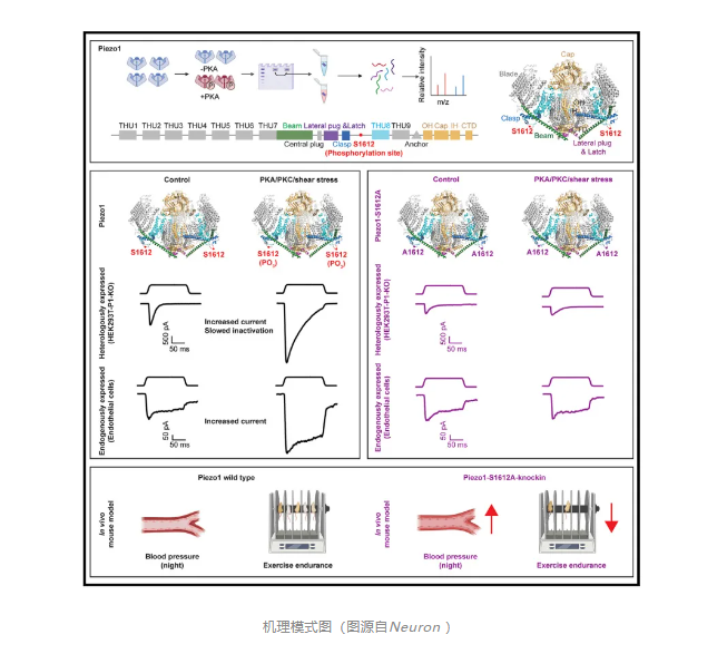
On September 12, 2024, Xiao Bailong of Tsinghua University and Ouyang Kunfu of Peking University jointly communicated inNeuron(IF=14.7)Published online entitled“Phosphorylation of Piezo1 at a single residue, serine-1612, regulates its mechanosensitivity and in vivo mechanotransduction function”The research paper,The study showedPhosphorylation of Piezo1 at a single residue, serine -1612, modulates its mechanosensitivity and mechanotransduction function in vivo.


Since the discovery of piezoproteins (including Piezo1 and Piezo2) as true mechanically activated cation channels in mammalian cells, Piezo1/2 has been found to mediate cellular mechanotransduction in a wide range of cell types. For example, endothelial Piezo1 plays a key role in sensing blood flow-related shear stress, which controls blood and lymphatic vessel development, vascular tone, and blood pressure regulation. Astrocyte Piezo1 is essential for monitoring the mechanical environment of the brain and contributes to adult neurogenesis and the structure and function of the brain. In a subset of sensory neurons, Piezo1 mediates mechanical pruritus.Piezo2 acts primarily as a major mechanoreceptor in sensory neurons and specialized sensory cells (e. g., Merkel cells), mediating light touch sensation, tactile pain, muscle stretch proprioception, sexual pleasure, lung inflation endoception, blood pressure fluctuations, bladder congestion, and gastrointestinal transit.

Structure-function studies of mouse Piezo1 (mPiezo1) and mPiezo2 revealed their 38-transmembrane (TM) topological folding characteristics of 2,547 and 2,822 amino acids, respectively, which trimerize to form a three-lobed, propeller-like channel and undergo a force-induced conformational transition from a highly bent state to a flattened state. The unique structural design and excellent deformability of Piezo1/2 may explain their superb mechanical sensitivity as multifunctional mechanotransduction channels in a variety of cell types and physiological processes.Despite significant progress in understanding the physiological importance and structure-function relationships of piezoelectric channels, how they can be post-translationally modified to fine-tune their channel properties to mediate their multifunctional mechanotransduction functions remains largely unexplored.
Activation of Piezo1 may trigger a cascade of downstream signaling components, including Ca 2 and protein kinases. For example, shear stress-induced activation of Piezo1 results in Ca 2 Influx and activation of cAMP-dependent protein kinase A (PKA), which then phosphorylates and activates endothelial cell nitric oxide (NO) synthesis (eNOS) to produce nitric oxide, a key vasodilator that causes vasodilation and blood pressure regulation. PKA is also known for mediating β-adrenergic regulation of electrical-mechanical coupling in the heart, with Piezo1 serving as the primary mechanotransduction channel for cardiomyocytes.In sensory dorsal root ganglion (DRG) neurons and xenogeneic expressing cell lines, piezo2-mediated mechanosensitive currents can be enhanced by PKA or protein kinase C (PKC) activation, which may be responsible for mechanical hyperalgesia or allodynia under inflammatory conditions. Although PKA and PKC mediate the regulation of piezoelectric channel function, it is currently unclear whether piezoelectric channels are directly phosphorylated by PKA or PKC at specific residues.
Here, the researchers found that PKA activation enhanced mechanosensitivity, slowed the kinetics of inactivation of mouse Piezo1, and identified a major phosphorylation site, serine -1612 (S1612), which also responds to PKC activation and shear stress. Mutant S1612 abrogates regulation of Piezo1 activity by PKA and PKC. Primary endothelial cells from Piezo1-S1612A knock-in mice lose PKA-and PKC-dependent phosphorylation and functional enhancement of Piezo1. Mutant mice exhibit activity-dependent increases in blood pressure and impaired exercise tolerance, similar to endothelial-specific Piezo1 knockout mice. Taken together, this study identified major PKA and PKC phosphorylation sites in Piezo1 and demonstrated their contribution to Piezo1-mediated physiological functions.