
Chongfan Technology
News
04
2026
-
08
The Xi’an Institute of Optics and Precision Mechanics has made progress in the field of label-free three-dimensional microscopy.
Author:
Recently, the research team led by Researcher Yao Baoli at the Laboratory of Transient Optics, Xi’an Institute of Optics and Precision Mechanics, Chinese Academy of Sciences, has made new advances in label-free three-dimensional microscopy. The related research findings, titled “Optical diffraction tomography using programmable LED array illumination and quantitative phase camera,” were published in the Journal of Physics: Photonics.
Optical diffraction tomography (ODT) is a label-free, non-invasive, quantitative three-dimensional microscopy technique that can reconstruct the three-dimensional refractive index distribution within transparent or semi-transparent samples, making it highly valuable in fields such as cell biology, histopathology, microbiology, and materials science. However, conventional ODT systems typically rely on Mach–Zehnder interferometer configurations, high-coherence laser sources, and mechanical scanning stages, resulting in complex setups with stringent stability requirements. Although intensity‑based ODT approaches leveraging LED arrays have emerged in recent years to simplify hardware, they generally require multiple intensity images and sophisticated iterative algorithms to retrieve phase information, thereby compromising imaging speed and reconstruction efficiency.
In response, the research team has developed an optical diffraction tomography method—LED‑ODT—that leverages a programmable LED array for illumination and a quantitative phase camera. This approach builds upon standard commercial microscopes: a programmable LED array enables rapid, non‑mechanical multi‑angle illumination, while the team’s self‑developed Q‑camera captures the sample’s complex amplitude information in a single exposure under a given illumination angle. Subsequently, by combining the Rytov approximation with the Fourier diffraction theorem, the multi‑angle complex amplitude data are mapped into a three‑dimensional frequency domain, ultimately reconstructing the sample’s three‑dimensional refractive index distribution (as shown in Figure 1).
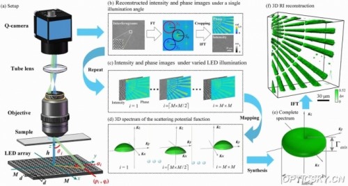
Figure 1. Schematic diagram of the LED-ODT system and its operating principle.
Based on the aforementioned LED‑ODT approach, the research team has developed a compact, label-free three-dimensional microscopy system comprising a 15×15 programmable LED array and a Q‑camera. This system achieves a maximum illumination angle of 52.9° and can acquire interference patterns across 225 illumination angles in just 2.7 seconds. The technology combines the complex amplitude measurement accuracy of conventional interferometric ODT, the phase‑measurement stability of common‑path ODT, and the hardware simplicity and operational convenience of intensity‑based ODT, thereby achieving an optimal balance among precision, stability, and ease of use.
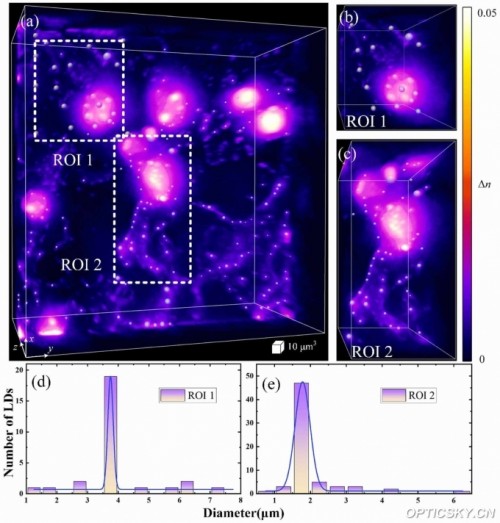
Figure 2. Three-dimensional refractive index reconstruction of COS-7 cells
Experimental validation demonstrates that this method delivers excellent three-dimensional refractive index imaging of both unstained COS-7 cells and water‑soaked Sterculia seeds: it not only resolves subcellular structures such as lipid droplets within COS-7 cells with high clarity (as shown in Figure 2), but also reveals the spatial heterogeneity of the three-dimensional refractive index distribution inside water‑soaked Sterculia seeds (as shown in Figure 3), thereby providing a new quantitative three-dimensional imaging approach for studying the structure and function of natural biomaterials.
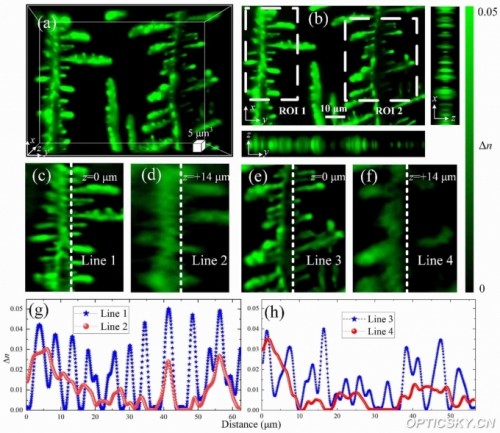
Figure 3. Three-dimensional refractive index reconstruction of water-soaked Sterculia seeds.
Li Yan explained, “Simply put, this technique captures a non-contact, full‑body 3D X‑ray image of microscopic samples—much like surrounding the sample with 225 miniature fill lights and sequentially illuminating it from different angles. There’s no need to move the instrument or reposition the sample; in just 2.7 seconds, it can reveal even the tiniest internal structures with crystal clarity. What’s more, it can be easily mounted onto a standard microscope, making it exceptionally accessible.”
The LED‑ODT system features a compact design, low cost, and ease of operation, enabling direct integration with standard optical microscopy platforms. While preserving imaging quality, it significantly enhances the practicality of ODT technology, offering a rapid, label‑free, high‑quality three‑dimensional quantitative microscopy tool for observing transparent biological samples, analyzing cellular metabolism, conducting histopathological research, and characterizing natural polymeric materials.
This research was supported by the National Natural Science Foundation of China, the National Key R&D Program, and the Chinese Academy of Sciences Youth Innovation Promotion Association, among other projects. The first author of the paper is Li Yan, a 2026‑enrolled doctoral student at the Xi’an Institute of Optics and Precision Mechanics; the corresponding authors are Researcher Min Junwei and Researcher Yao Baoli. The Xi’an Institute of Optics and Precision Mechanics served as both the primary completing institution and the corresponding institution.
Source: Xi’an Institute of Optics and Precision Mechanics, Chinese Academy of Sciences
LATEST NEWS
Thank you for visiting the official website of Chongfan Technology. If you have cooperation intentions or suggestions, please contact us through the following methods, and we will reply as soon as possible, thank you!
Address: Room 403, Building 6, Phase III of R&D, No. 36 Xiyong Avenue, High tech Zone, Chongqing, China.
Telephone: +86-13658337211
E-mail: Sales@cfkeji.net
Website: www.cfkeji.net
Mobile Version